Confocal Microscopy
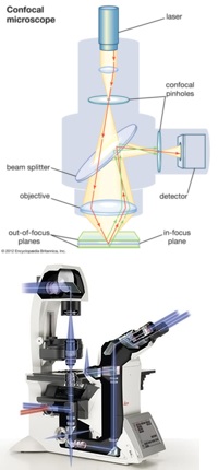
Confocal Imaging Concept The primary functions of a confocal microscope are to produce a point source of light and reject out-of-focus light, which provides the ability to image deep into tissues with high resolution, and optical sectioning for 3D reconstructions of imaged samples. The basic principle of confocal microscopy is that the illumination and detection optics are focused on the same diffraction-limited spot, which is moved over the sample to build the complete image on the detector. While the entire field of view is illuminated during confocal imaging, anything outside the focal plane contributes little to the image, lessening the haze observed in standard light microscopy with thick and highly-scattering samples, and providing optical sectioning.
Manual inverted microscope (Model: Leica DMi8) with TCS SPE confocal module. It is designed for all common microscope applications and techniques. The microscope equipped with traditional transmitted light, fluorescence system, three laser lines (488nm 532nm 635nm) and tunable emission detector. System is equipped with LAS X software that allows to process and quantify the acquired images.
